
3D cranial anthropometry: cranial analysis from a three-dimensional scan
How 3D cranial anthropometry calculates CVAI, cephalic index and 3D asymmetry from a scan, and what makes measurements reliable across assessments.
3D cranial anthropometry uses a head scan to reconstruct the cranial surface and obtain objective measurements of cranial shape. When acquisition and landmark placement are standardized, the method can increase the reproducibility of measurements and provides a three-dimensional record of cranial morphology.
Beyond traditional indices such as CVAI and the cephalic index, three-dimensional analysis makes it possible to assess how volume is distributed across the regions of the head and to document how cranial shape changes over the course of treatment.
How it works
The process starts by capturing the surface of the head with a 3D scanner. The result is a mesh that represents the external geometry of the skull.
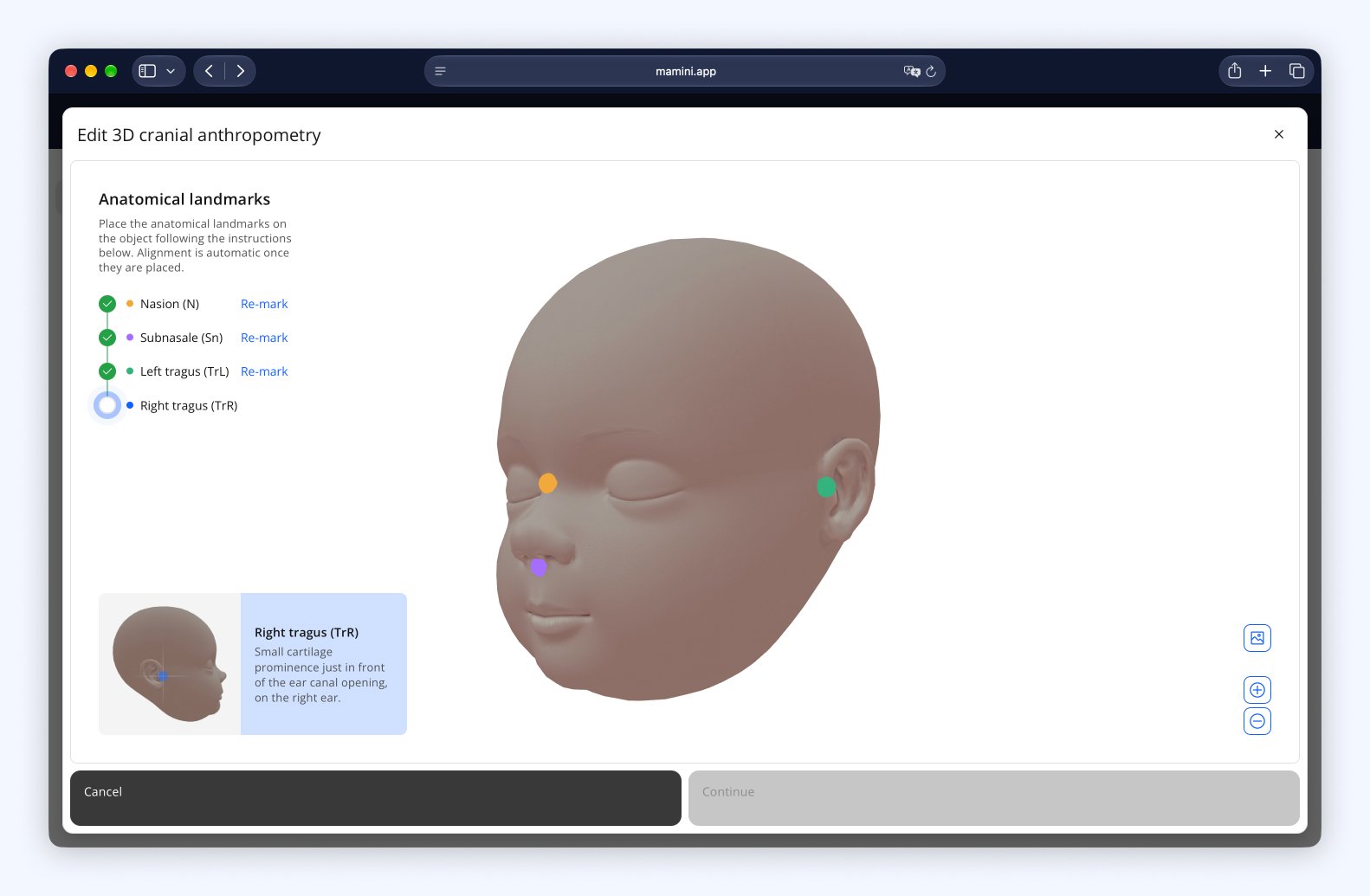
On this reconstruction, the clinician places anatomical landmarks that orient the head. In the model described by Linz et al. (2022), there are four landmarks: the nasion, the subnasale and the two tragi. Because the orientation comes from the anatomy, it removes the need to level the head manually and does not depend on how the infant was positioned during the scan.
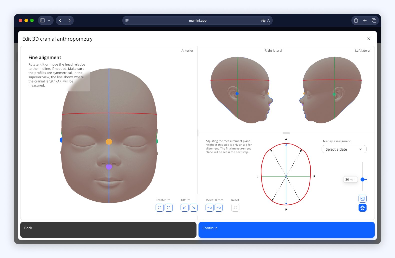
Once the head is oriented, the measurement plane is defined: the horizontal section where the measurements are taken. The most common conventions are a plane 30 mm above the ear line and the plane of the largest head circumference. More important than the convention chosen is repeating the same plane across the patient's assessments, so that the results are comparable.
From there, the system calculates the measurements and the indices.
What 3D measures beyond CVAI and the cephalic index
Conventional cranial anthropometry relies mainly on linear measurements taken between specific anatomical landmarks. Three-dimensional analysis extends that assessment by using the geometry of the cranial surface to obtain further parameters.
The measurements that can be obtained include:
CVAI: quantifies the asymmetry between oblique diagonals of the cranial vault.
Cephalic index: relates the width of the skull to its anteroposterior length.
ACAI: quantifies volumetric asymmetry in the anterior region of the skull.
PCAI: quantifies volumetric asymmetry in the posterior region of the skull.
3D asymmetry index: assesses asymmetry taking the three-dimensional surface of the head into account.
Head circumference: can be derived from the three-dimensional mesh.
Other geometric measurements: can be calculated from the reconstruction of the cranial surface.
These measurements complement the clinical assessment and the traditional indices, allowing a broader analysis of cranial morphology.
Anterior and posterior asymmetry
ACAI and PCAI make it possible to quantify volume asymmetry in different regions of the head: ACAI in the anterior region, PCAI in the posterior region. Together, the indices help identify how the volume difference is distributed between those regions.
This information complements CVAI, which focuses on the difference between the diagonals of the cranial vault.
3D asymmetry index
The 3D asymmetry index, proposed by Linz et al. (2022), quantifies cranial asymmetry from the distribution of volume across the four quadrants of the head, identified as Q1, Q2, Q3 and Q4:
3D asymmetry index = |ln((Q4 × Q2) / (Q3 × Q1))|
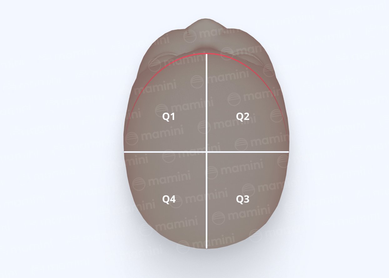
The calculation compares the product of the volumes of diagonally opposite quadrants. When the volumes are distributed symmetrically, the index approaches 0; the greater the difference between the sides, the higher the value tends to be.
Unlike CVAI, which uses linear measurements, the 3D asymmetry index considers how volume is distributed across the whole head. Linz et al. showed that this parameter performed well in identifying and classifying plagiocephaly in their sample, representing three-dimensional asymmetry in a way that complements traditional measurements.
Head circumference and z-score
Head circumference can be obtained directly from the three-dimensional reconstruction. Using the patient's age and sex, the value is compared with the World Health Organization reference curves, with corrected age for preterm infants, to obtain the z-score.
Head circumference and its z-score are complementary to the analysis of cranial shape. They do not replace the assessment of the asymmetry indices or of the cephalic index.
Benefits of 3D cranial anthropometry
Three-dimensional assessment
The 3D model represents the cranial surface in three dimensions, making it possible to visualize and analyze features that isolated linear measurements do not represent, such as how volume is distributed across the regions of the head.
Reproducibility
Orientation comes from anatomical landmarks and the measurement plane is defined mathematically, which reduces variation related to positioning and to defining the sections manually. With standardized landmark placement and the same plane across visits, serial assessments tend to be more consistent.
Fast acquisition without prolonged direct contact
Depending on the equipment used, the scan can be performed quickly and without prolonged contact with the infant's head, which can make acquisition easier in small patients.
A permanent record of morphology
Beyond the numbers, the scan produces a three-dimensional representation of the cranial surface. This record makes it possible to document the shape of the head at a given moment and compare it with later assessments.
Visible progress
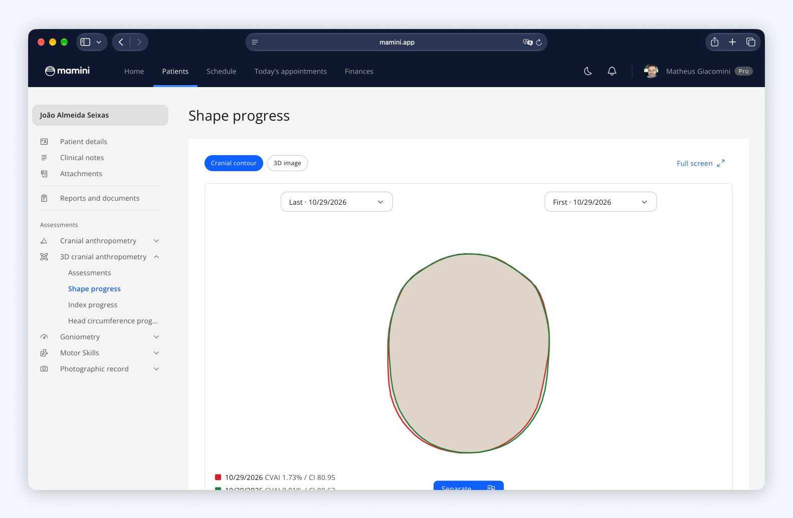
With repeated assessments, the contour of the head from two dates can be overlaid to show, at the same scale, where the shape has changed. This image helps the clinician follow the response to treatment and show progress to the family, for whom a shape often says more than an index.
Comparing manual and 3D cranial anthropometry
Manual and 3D cranial anthropometry can both be used to quantify cranial shape, but they have different methodological characteristics.
Manual cranial anthropometry uses instruments and landmarks defined by the examiner. It is accessible, can be performed quickly and has good clinical utility when the technique is standardized.
3D cranial anthropometry uses a digital reconstruction of the cranial surface. Beyond linear measurements, it allows volume analysis and a record of three-dimensional morphology.
A CVAI calculated from a 3D section and a CVAI obtained by manual cranial anthropometry can show similar trends, but they should not be treated as identical measurements. Differences in acquisition, positioning, landmark definition and calculation method can influence the results. For serial follow-up, it is therefore important to keep the same method and measurement protocol whenever possible.
Precautions and limitations
The quality of the 3D analysis depends directly on the quality of the acquisition. Movement by the infant, missing regions of the surface, hair, accessories, poor positioning or reconstruction failures can all interfere with the measurements.
Landmark placement can also introduce variation. For this reason, automating certain steps does not remove the need for an acquisition protocol and for the clinician's review.
Values from different devices or software may not be directly comparable without method validation and standardized protocols. Quantitative analysis should therefore be interpreted together with the clinical examination and the individual course of the child.
3D cranial anthropometry in follow-up
The main advantage of 3D scanning lies not only in a detailed assessment at a given moment, but in the ability to document progress. By repeating assessments throughout treatment, the clinician can compare:
CVAI and cephalic index over time;
anterior and posterior asymmetry;
the 3D asymmetry index;
head circumference and its z-score;
changes in the shape of the head;
the response to the interventions carried out.
Using 3D in cranial remolding orthosis assessment
3D scanning is widely used in the assessment and planning of cranial remolding orthoses, making it possible to document the shape of the head and provide data for the planning and custom manufacture of the device.
The same technology can also be used long before that stage, in routine follow-up, to measure, classify and document the progress of infants under conservative management.
Using 3D does not, on its own, determine whether an orthosis is needed. The indication should take into account the clinical assessment, the severity of the deformity, the child's age, the course of the condition and the response to conservative measures.
How Mamini does it
In Mamini, 3D cranial anthropometry is part of the patient record. The clinician uploads the scan and, in three steps, places the four anatomical landmarks, checks the alignment of the head and chooses the measurement plane. The system warns when a landmark appears to be out of place and allows the plane from the previous assessment to be repeated, so that assessments remain comparable.
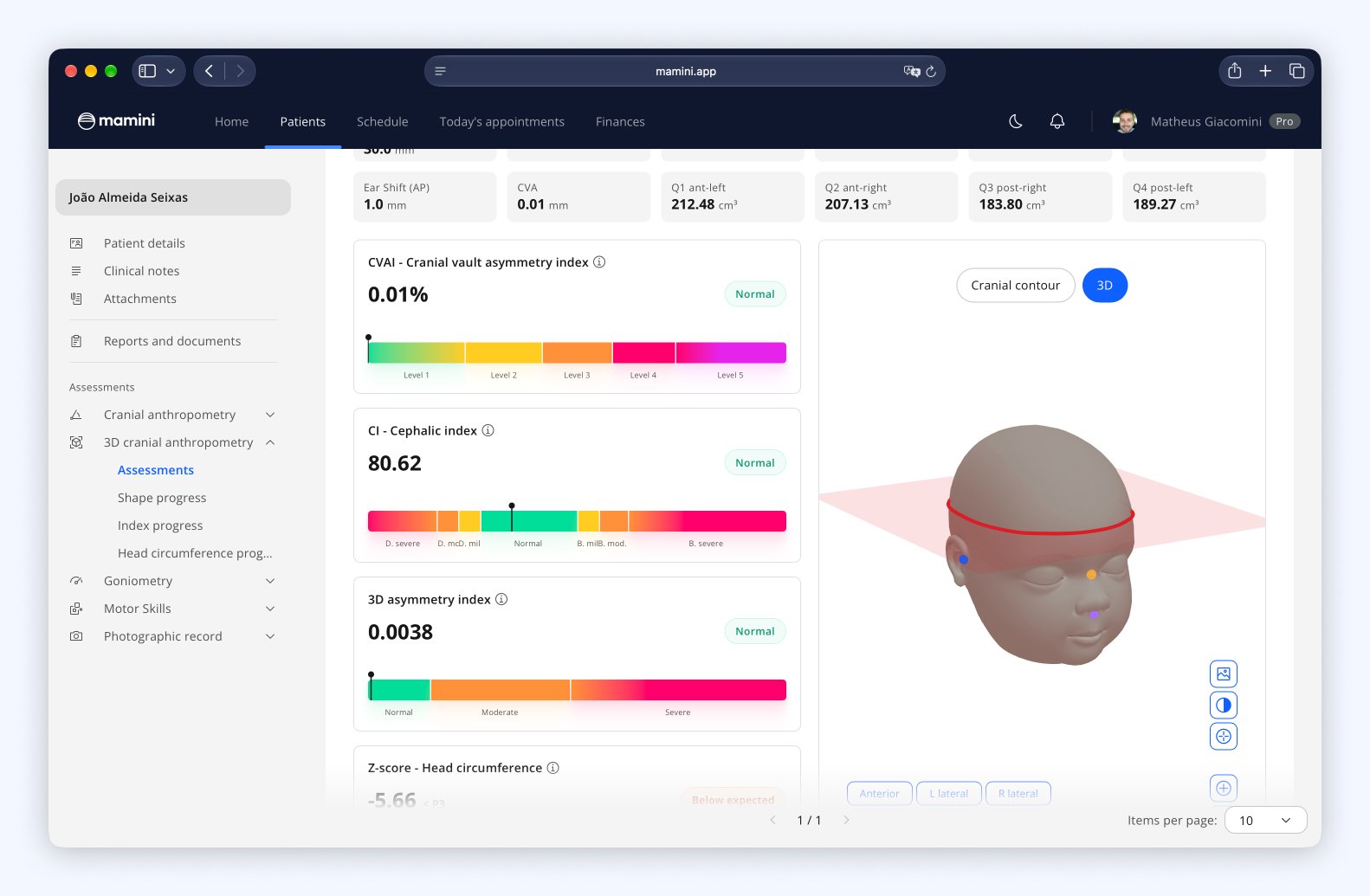
The results include CVAI, the cephalic index, the 3D asymmetry index, head circumference with z-score and ear shift, and the clinician chooses the CVAI formula and the classification scale they use.
In follow-up, charts show how the indices and head circumference change over time, and shape progress overlays the contours of two assessments at the same scale. The PDF report brings together measurements, indices and images.
Because Mamini does not charge per assessment, unlike the common practice in this market, scanning can be part of routine follow-up and not just a one-off assessment.
References
Linz C et al. Using a 3D asymmetry index as a novel form for capturing complex three-dimensionality in positional plagiocephaly. Sci Rep. 2022;12:20831. DOI: 10.1038/s41598-022-24555-1
Holowka MA et al. Plagiocephaly Severity Scale to Aid in Clinical Treatment Recommendations. J Craniofac Surg. 2017;28(3):717-722. DOI: 10.1097/SCS.0000000000003520
Graham JM Jr et al. Deformational brachycephaly in supine-sleeping infants. J Pediatr. 2005;146(2):253-257. DOI: 10.1016/j.jpeds.2004.10.017
Likus W et al. Cephalic index in the first three years of life: study of children with normal brain development based on computed tomography. ScientificWorldJournal. 2014;2014:502836. DOI: 10.1155/2014/502836
WHO Multicentre Growth Reference Study Group. WHO Child Growth Standards: Head circumference-for-age, arm circumference-for-age, triceps skinfold-for-age and subscapular skinfold-for-age: Methods and development. Geneva: World Health Organization; 2007.
Congress of Neurological Surgeons. Evidence-Based Guidelines: The Role of Imaging. Guidelines for the Management of Patients with Positional Plagiocephaly.
Frequently asked questions
What is 3D cranial anthropometry?
It is the quantitative analysis of cranial shape from a three-dimensional reconstruction of the head surface. The method makes it possible to obtain linear measurements, asymmetry indices and other geometric parameters.
What are the benefits of 3D cranial anthropometry?
3D cranial anthropometry makes it possible to assess the head in three dimensions, including the distribution of volume, with measurements standardized by anatomical landmarks. The scan is fast, does not require prolonged contact with the infant and creates a permanent record of cranial shape that can be compared across assessments.
Does 3D cranial anthropometry replace clinical assessment?
No. 3D scanning provides objective measurements and documentation of cranial shape, but the results should be interpreted together with the clinical examination, the child's age, the course of the condition and the other aspects of the assessment.
Is the CVAI from 3D the same as the CVAI from manual cranial anthropometry?
Both can use the same geometric logic, based on the difference between the cranial diagonals, but the results should not automatically be treated as identical measurements. The acquisition method, positioning and landmark definition can all influence the result.
Is 3D scanning more accurate than manual cranial anthropometry?
It would not be appropriate to claim that one method is universally more accurate. 3D cranial anthropometry offers advantages related to documenting the cranial surface, geometric analysis and standardizing certain steps. The quality of the result depends on the acquisition, the processing and the protocol used.
Can 3D scanning be used to follow treatment?
Yes. Repeating the scan makes it possible to compare measurements and three-dimensional representations over time, helping to document the progression of cranial shape and the response to interventions.
Does Mamini indicate whether a cranial remolding orthosis is needed?
Mamini provides measurements and objective documentation of cranial shape. The indication for an orthosis should be made by the clinician responsible, considering the clinical assessment as a whole, the child's age, the severity, the course of the condition and the response to treatment.