
How to calculate CVAI and the cephalic index in cranial anthropometry
A step-by-step guide to calculating CVAI and the cephalic index: the cranial anthropometry measurements (cranial length, cranial width and diagonals), the formulas, the classification (CHOA scale and CI ranges) and clinical interpretation.
Quantifying cranial asymmetry is what turns a clinical impression into objective follow-up. Among the main indices used in cranial anthropometry are CVAI (cranial vault asymmetry index) and the cephalic index (CI). This guide covers the required measurements, the formulas, the classification and the precautions for keeping assessments consistent.
The cranial anthropometry measurements
Using a caliper and standardized landmarks, collect:
Measurement | Description |
|---|---|
Cranial length (AP) | Anteroposterior diameter (length) |
Cranial width (ML) | Biparietal diameter (width) |
Diagonal A | Left oblique diagonal |
Diagonal B | Right oblique diagonal |
HC | Head circumference (optional) |
Diagonals A and B are measured at approximately 30° from the midline, between frontal landmarks and contralateral occipital landmarks. Standardizing the landmarks and keeping the technique consistent across sessions are essential so that the changes observed reflect the real progression of cranial shape, and not just measurement variation.
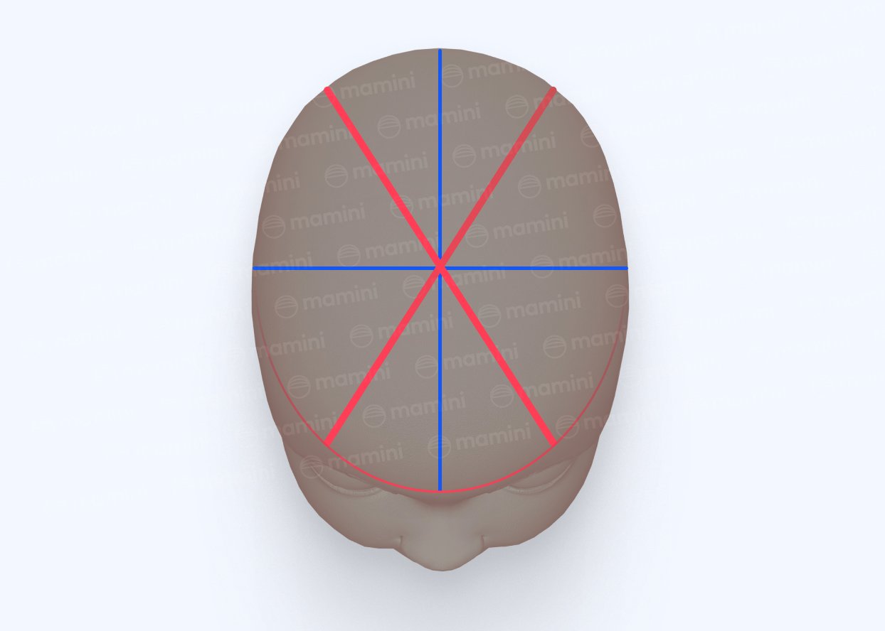
CVAI: cranial vault asymmetry index
CVAI quantifies the asymmetry between the two oblique diagonals of the skull.
Mamini's default formula is:
CVAI = |Diagonal A − Diagonal B| / longer diagonal × 100
In other words, the absolute difference between the two diagonals is divided by the longer one and multiplied by 100.
The literature describes different ways of calculating CVAI, which differ mainly in the denominator used. In CVAI(S), for example, the difference is divided by the shorter diagonal (Loveday and de Chalain, 2001). In this article, we use the formula from the CHOA (Children's Healthcare of Atlanta) severity scale, in which the difference between the diagonals is divided by the longer diagonal. What matters most, especially in clinical follow-up, is using the same method consistently across assessments.
Example: Diagonal A = 130 mm and Diagonal B = 140 mm
CVAI = |130 − 140| / 140 × 100 = 7.14%
In this example, the result corresponds to the moderate classification.
CVAI classification
Mamini's default scale is the CHOA severity classification:
Classification | CVAI |
|---|---|
Normal | < 3.5% |
Mild | ≥ 3.5% and < 6.25% |
Moderate | ≥ 6.25% and < 8.75% |
Severe | ≥ 8.75% and ≤ 11.0% |
Very severe | > 11.0% |
Another scale in use is that of Kim et al. (2024), defined with CVAI(S): normal below 3.5%, mild from 3.5% to 6%, moderate above 6% up to 10%, and severe above 10%. In Mamini, the formula and the scale can be chosen in each clinician's settings.
Cephalic index (CI)
The cephalic index relates the width of the skull to its length and is used to characterize cranial proportion, including brachycephalic and dolichocephalic patterns.
The formula is:
CI = cranial width / cranial length × 100
The index is a ratio between the width and length measurements, multiplied by 100.
Example: cranial width = 130 mm and cranial length = 138 mm
CI = 130 / 138 × 100 = 94.2
In this example, the result corresponds to moderate brachycephaly, according to the ranges adopted by Mamini.
Cephalic index classification
Mamini uses the following classification ranges:
Classification | CI |
|---|---|
Severe dolichocephaly | < 70 |
Moderate dolichocephaly | 70 to < 73 |
Mild dolichocephaly | 73 to < 76 |
Normal | 76 to < 90 |
Mild brachycephaly | 90 to < 93 |
Moderate brachycephaly | 93 to < 97 |
Severe brachycephaly | ≥ 97 |
These ranges combine specific references for classifying the different patterns of cranial proportion, according to the sources used by Mamini.
Reproducibility: the most common pitfalls
Standardize the landmarks. The same anatomical landmarks should be used in every assessment. Small differences in where the diagonals are placed can change the CVAI.
Keep the same technique across sessions. To follow progress reliably, it is important to repeat the same measurement method throughout treatment.
Manual and 3D cranial anthropometry: what you gain in indices
CVAI and the cephalic index can be obtained either by manual cranial anthropometry, with a caliper, or by 3D cranial anthropometry, from a head scan.
Besides allowing greater standardization of acquisition and analysis, 3D cranial anthropometry makes it possible to assess aspects of cranial shape that linear measurements alone do not represent.
These include:
Quadrant asymmetry indices, anterior (ACAI) and posterior (PCAI): help identify where the asymmetry is most concentrated, not just its overall magnitude.
3D asymmetry index: assesses the deformation of the cranial surface considering its three-dimensional shape, not just the diagonals.
Mesh-derived head circumference and ear shift: add further quantitative information to the assessment of cranial morphology.
In practice, manual cranial anthropometry is an accessible way to follow CVAI and the cephalic index. 3D analysis extends this assessment by allowing a more detailed characterization of cranial shape.
Read also: 3D cranial anthropometry: cranial analysis from a three-dimensional scan
How Mamini does it for you
In Mamini, you can use both assessment methods: manual cranial anthropometry, by entering the measurements, and 3D cranial anthropometry, from a scan.
In both, CVAI and the cephalic index are calculated and classified automatically, using the formula and scale chosen by the clinician. The calculation is kept consistent across assessments, making it easier to follow the patient's progress.
Results are stored in the patient's history, can be followed in charts and are used to generate the report.
References
Holowka MA et al. Plagiocephaly Severity Scale to Aid in Clinical Treatment Recommendations. J Craniofac Surg. 2017;28(3):717-722. DOI: 10.1097/SCS.0000000000003520 (CHOA scale)
Graham T et al. Significant Factors in Cranial Remolding Orthotic Treatment of Asymmetrical Brachycephaly. J Clin Med. 2020;9(4):1027. DOI: 10.3390/jcm9041027 (CVAI level names)
Loveday BP, de Chalain TB. Active counterpositioning or orthotic device to treat positional plagiocephaly? J Craniofac Surg. 2001;12(4):308-313. DOI: 10.1097/00001665-200107000-00003 (CVAI(S))
Kim J et al. Effectiveness of Helmet therapy for infants with moderate to severe positional plagiocephaly. Clin Exp Pediatr. 2024;67(1):46-53. DOI: 10.3345/cep.2023.00626 (Kim scale)
Graham JM Jr et al. Deformational brachycephaly in supine-sleeping infants. J Pediatr. 2005;146(2):253-257. DOI: 10.1016/j.jpeds.2004.10.017 (brachycephaly)
Choi H et al. Outcome Analysis of the Effects of Helmet Therapy in Infants with Brachycephaly. J Clin Med. 2020;9(4):1171. DOI: 10.3390/jcm9041171 (brachycephaly)
Cohen MM Jr, MacLean RE, eds. Craniosynostosis: Diagnosis, Evaluation, and Management. 2nd ed. New York: Oxford University Press; 2000. (dolichocephaly)
Likus W et al. Cephalic index in the first three years of life: study of children with normal brain development based on computed tomography. ScientificWorldJournal. 2014;2014:502836. DOI: 10.1155/2014/502836 (dolichocephaly)
Frequently asked questions
In CVAI, is the denominator the longer or the shorter diagonal?
Mamini's default is the longer diagonal: the difference between the two diagonals is divided by the longer one, following the formula used in the CHOA scale. Other formulas described in the literature, such as CVAI(S), use the shorter diagonal as the denominator, and Mamini also lets you choose it in settings. It is therefore important to identify which method is being used and to keep the same criterion when comparing assessments of the same patient.
How can I make sure progress between sessions is reliable?
Standardize the landmarks, the measurement technique and the calculation method, repeating them the same way in every assessment.
What additional indices does 3D cranial anthropometry offer?
Beyond CVAI and the cephalic index, 3D analysis makes it possible to assess quadrant asymmetry, anterior (ACAI) and posterior (PCAI), the 3D asymmetry index, mesh-derived head circumference and ear shift. These measurements complement the assessment of linear indices and allow a more detailed analysis of cranial morphology.