
Positional cranial asymmetry: clinical assessment, cranial anthropometry and management
A clinical guide to cranial asymmetry in infants: classification, differential diagnosis with craniosynostosis, quantification by cranial anthropometry (CVAI and cephalic index) and follow-up.
Deformational cranial asymmetry is a common reason for assessment in the first months of life. An appropriate approach involves recognizing the pattern of the deformity, distinguishing it from changes related to craniosynostosis, identifying associated factors such as positional preference and congenital muscular torticollis, and quantifying the asymmetry objectively and reproducibly.
Cranial anthropometry turns the assessment of cranial shape into measurements that can be compared over time, helping the clinician document progress and make clinical decisions.
Definition and associated factors
Deformational plagiocephaly and brachycephaly result from external forces acting on a developing skull, whose sutures remain open and allow cranial growth. Sustained head positioning, positional preference and associated conditions, such as congenital muscular torticollis, can contribute to the development or persistence of the deformity.
The frequency of these changes increased after the recommendation that infants sleep on their backs, a measure that is essential for reducing the risk of sudden infant death syndrome and that should be maintained.
The severity of the deformity and its course depend on several factors, including age, rate of cranial growth, positioning pattern, cervical mobility and duration of exposure to pressure. Early assessment therefore makes it possible to identify modifiable factors and follow the response to the measures adopted.
Clinical classification
Deformational patterns
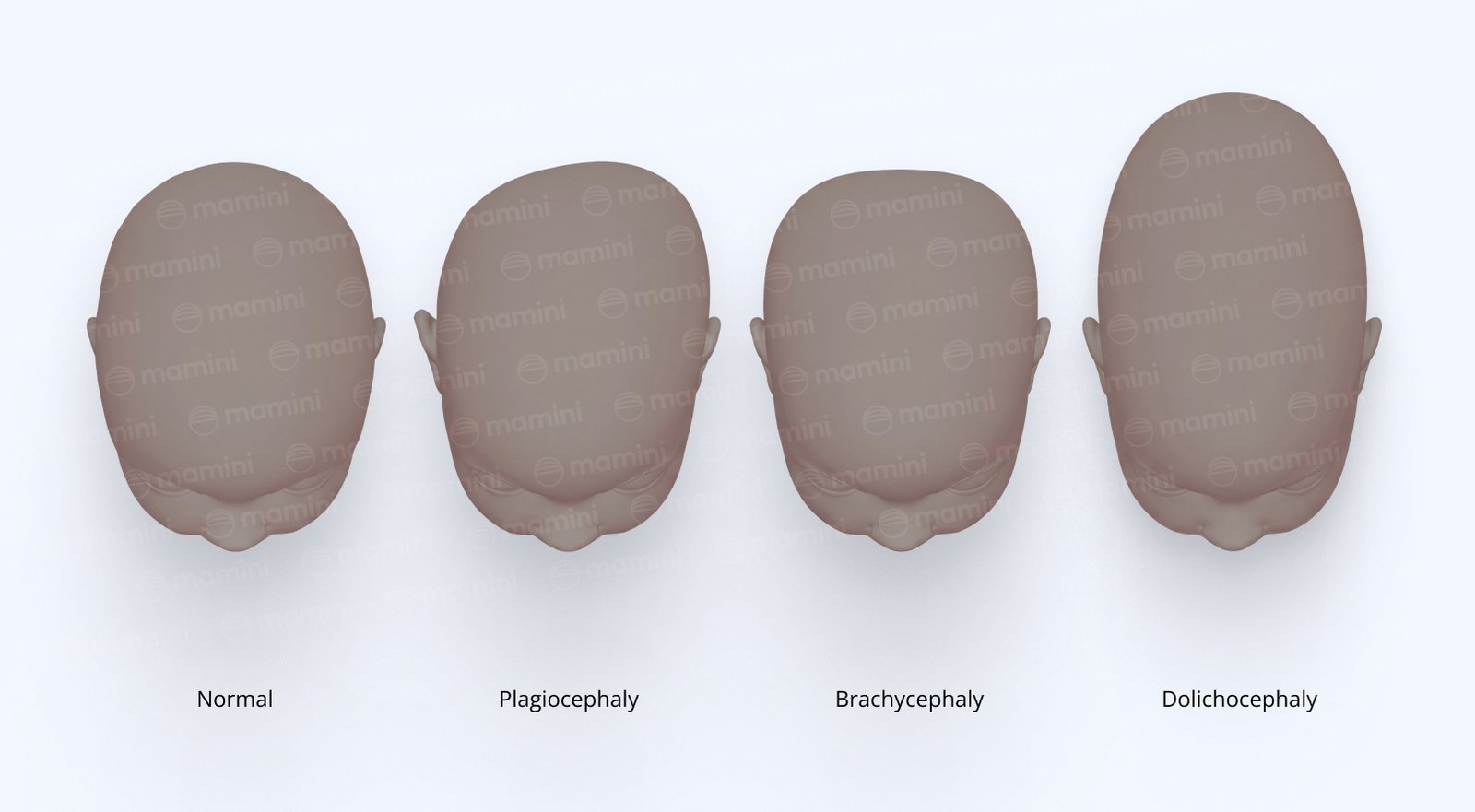
Plagiocephaly: predominantly unilateral occipital flattening. Seen from above, the skull tends to take on a parallelogram-like shape, often accompanied by anterior displacement of the ear on the same side and ipsilateral frontal bossing. There may be associated facial asymmetry.
Brachycephaly: predominantly symmetrical occipital flattening, with a relative reduction in anteroposterior length and a proportional increase in cranial width, producing a shorter, wider skull.
Deformational dolichocephaly or scaphocephaly: a relatively long, narrow skull. It may be seen, among other situations, in some preterm infants kept in certain positions in bed for long periods.
Clinical classification should consider the overall shape of the skull, the position of the ears, the frontal region, the cranial base and any facial changes. Visual assessment is important, but it can be complemented by objective measurements to quantify the deformity and follow its course.
Severity scales
In clinical assessment, the Argenta classification can be used to characterize different patterns of plagiocephaly and brachycephaly.
For objective quantification of plagiocephaly, one of the classifications used is the severity scale based on the Cranial Vault Asymmetry Index (CVAI), developed by Children's Healthcare of Atlanta (CHOA).
Classification | CVAI |
|---|---|
Normal | < 3.5% |
Mild | ≥ 3.5% and < 6.25% |
Moderate | ≥ 6.25% and < 8.75% |
Severe | ≥ 8.75% and ≤ 11.0% |
Very severe | > 11.0% |
Source: Holowka et al. (2017). By default, Mamini classifies CVAI using these ranges; the Kim et al. (2024) scale can also be selected in settings.
These cutoffs correspond to Mamini's default scale and should be interpreted together with the clinical examination, the child's age, the pattern of the deformity and its course. CVAI alone does not determine treatment.
Differential diagnosis: deformational vs. synostotic
Distinguishing a deformational cranial deformity from craniosynostosis is one of the most important steps of the assessment, because the conditions have different mechanisms and management.
Some clinical findings can help with this distinction:
Ear position: in posterior deformational plagiocephaly, the ear on the flattened side tends to be displaced anteriorly. In lambdoid craniosynostosis, it may be displaced posteriorly and inferiorly, together with a more trapezoidal skull shape.
Skull shape: deformational plagiocephaly usually has a parallelogram-like configuration seen from above, whereas lambdoid synostosis may produce a more trapezoidal shape.
Mastoid region: mastoid bulging may be present in lambdoid craniosynostosis.
Suture palpation: a bony ridge or a suspicious change along a suture may raise the hypothesis of craniosynostosis.
Course: persistence or progression of the deformity despite properly conducted measures may justify further investigation.
These findings should not be used in isolation to establish the diagnosis. When there are signs suggestive of craniosynostosis or diagnostic doubt, specialist assessment is indicated. The need for and type of imaging should be defined according to the clinical assessment.
Quantification: cranial anthropometry and indices
Cranial anthropometry makes it possible to quantify features of cranial shape through standardized measurements. When performed consistently, it allows different assessments to be compared and progress to be documented objectively.
The parameters used include the cranial vault asymmetry index (CVAI) and the cephalic index (CI).
Cranial vault asymmetry index (CVAI)
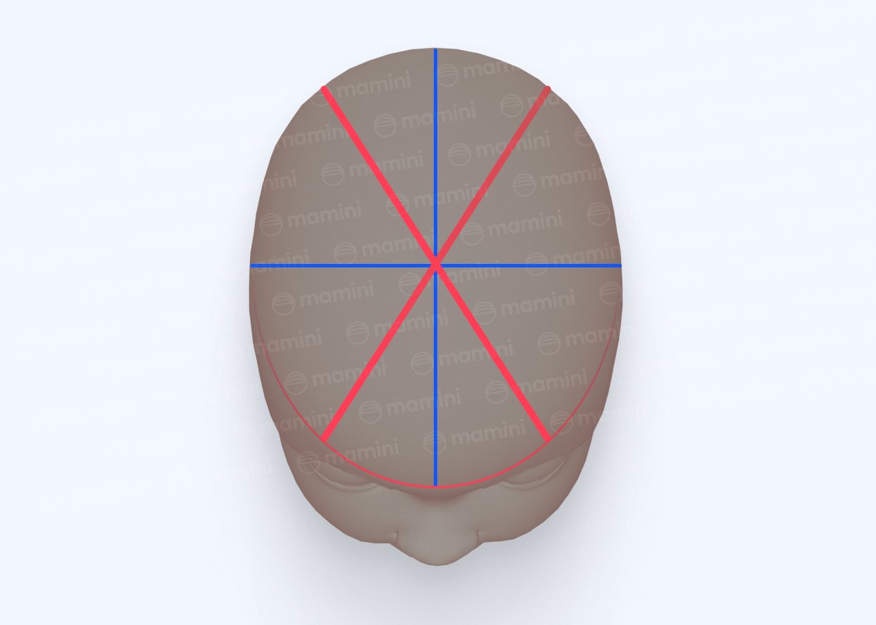
CVAI quantifies the difference between two oblique diagonals of the cranial vault.
Considering the left (Diagonal A) and right (Diagonal B) oblique diagonals, Mamini's default calculation is:
CVAI = |Diagonal A − Diagonal B| / longer diagonal × 100
The denominator is the longer of the two diagonals.
The result expresses, in relative terms, the difference between the diagonals. The higher the value, the greater the oblique asymmetry measured.
CVAI is particularly useful in assessing plagiocephaly, but it can be complemented by other measurements, by clinical assessment and, when available, by three-dimensional analysis of the cranial surface.
Cephalic index (CI)
The cephalic index relates cranial width to anteroposterior length:
CI = cranial width / cranial length × 100
where:
Cranial width (ML): biparietal diameter.
Cranial length (AP): anteroposterior diameter.
The cephalic index helps characterize wider and shorter skull patterns, such as brachycephaly, or longer and narrower ones, such as dolichocephaly.
As a ratio between two measurements multiplied by 100, the index is dimensionless and can exceed 100 in cases of marked brachycephaly.
For classification in Mamini, the following operational ranges are used:
Severe dolichocephaly: < 70.
Moderate dolichocephaly: 70 to < 73.
Mild dolichocephaly: 73 to < 76.
Normal: 76 to < 90.
Mild brachycephaly: 90 to < 93.
Moderate brachycephaly: 93 to < 97.
Severe brachycephaly: ≥ 97.
These ranges should be understood as classification criteria used by the system, not as a universal classification applicable regardless of age, population, measurement method and reference used.
Measurement methods
Manual cranial anthropometry: uses instruments such as a caliper, measuring tape or plagiocephalometer. It is accessible and can show good reproducibility when the anatomical landmarks, positioning and measurement technique are standardized.
3D scanning: reconstructs the cranial surface and obtains measurements from the three-dimensional model. It can increase the standardization and reproducibility of serial assessments, provided acquisition and processing are performed consistently.
Standardized photographic record: visually documents progress and complements the quantitative measurements, making communication between clinician and family easier.
In Mamini, CVAI, the cephalic index and other measurements can be calculated from manual cranial anthropometry or from three-dimensional analysis. Results are organized to make it easier to compare assessments, follow progress and generate clinical documentation.
Standardizing acquisition and measurement is essential. Differences in positioning, anatomical landmarks or technique can influence the results, regardless of the method used.
Read also: How to calculate CVAI and the cephalic index in cranial anthropometry and 3D cranial anthropometry: cranial analysis from a three-dimensional scan
Associated assessment of congenital muscular torticollis
Congenital muscular torticollis and deformational cranial asymmetry may be associated. Limited cervical mobility and a preference for keeping the head in a certain position can increase the exposure of one region of the skull to pressure and help maintain the deformity.
For this reason, when torticollis or positional preference is suspected, assessing cervical mobility is important. Depending on the case, the range of cervical rotation and lateral flexion, the presence of postural asymmetries and the infant's ability to actively turn the head to both sides should be considered.
Identifying a cervical limitation may point to the need for specific intervention and should be considered together with the assessment of cranial shape.
Read also: Congenital muscular torticollis: assessment by goniometry and management
Management by severity and age
Management should be individualized, considering age, severity of the deformity, course, associated factors and response to the measures adopted. There is no single treatment indicated for all cases.
Repositioning and parent guidance
Repositioning is one of the main conservative measures, especially in the first months of life.
Guidance may include:
alternating the head position during sleep, always keeping the infant on their back to sleep;
visual and auditory stimulation from the side opposite the flattening during awake periods;
more time in different positions while the infant is awake and supervised;
tummy time while awake and supervised;
reducing unnecessary time in devices that keep prolonged pressure on the occipital region;
guiding the family on safe ways to encourage symmetrical movements.
The goal is not to change the safe sleep recommendation, but to reduce prolonged pressure on the same region of the skull while the infant is awake.
Physical therapy
Physical therapy is especially relevant when there is congenital muscular torticollis, limited cervical mobility or a persistent positional preference.
The approach may include range-of-motion exercises, encouraging active and symmetrical movements, strengthening and postural control, positioning strategies and caregiver education.
The need for and intensity of treatment should be defined according to the individual assessment. The progression of cervical mobility and cranial asymmetry can be followed across visits to verify the response to the intervention.
Manual therapy
Manual techniques may be used as part of an individualized physical therapy approach, especially when there is restricted mobility or associated musculoskeletal changes.
When used in infants, they should be performed by a trained professional, with techniques appropriate to the child's age and clinical condition. Manual therapy should not be presented on its own as a treatment for cranial deformity, nor replace measures with stronger support for managing torticollis, such as therapeutic exercise, encouraging active movement, positioning and caregiver education.
Different professionals may use specific manual approaches. The choice of technique should consider the clinical assessment and the professional's training, without assuming that any specific approach is superior to the others in correcting cranial asymmetry.
Cranial orthosis
A cranial orthosis may be considered in certain cases of deformational plagiocephaly or brachycephaly, especially when the deformity is moderate to severe, persists despite properly conducted conservative measures or presents at an older age, when the potential for spontaneous remodeling is lower.
The decision should mainly consider:
severity of the deformity;
the child's age;
the course of the measurements;
response to repositioning and physical therapy;
presence of associated factors;
expected cranial growth;
the assessment of the responsible clinician.
In general, when indicated, an orthosis tends to give better results when used during the period of fastest cranial growth. However, there is no single age or severity cutoff that determines its indication for every child.
The evidence on orthoses is not uniform, especially in moderate cases. Studies show different results depending on the population, age, severity and criteria used. The indication should therefore be individualized and not based solely on a CVAI or cephalic index value.
Follow-up and documentation
Serial measurement makes it possible to distinguish clinical impression from objectively documented progress.
Recording CVAI, the cephalic index and, when relevant, other measurements across visits makes it possible to:
follow the progression of cranial shape;
verify the response to interventions;
identify absent or slowing improvement;
support decisions on continuing or changing management;
document treatment objectively;
make communication among the professionals involved easier.
A standardized photographic record can complement the quantitative data and help show changes in cranial shape over time.
This is the follow-up Mamini was built to make easier. The clinician records manual or 3D cranial anthropometry, obtains the indices in a standardized way and follows the results in the history and in charts, with report generation and documentation of the assessments.
When to refer
Specialist assessment should be considered especially in the following scenarios:
clinical signs suggestive of craniosynostosis, such as a suspicious change along a suture, a bony ridge, an atypical cranial pattern or a course incompatible with a positional deformity;
diagnostic doubt between a positional deformity and craniosynostosis;
significant or progressive asymmetry;
lack of response to properly conducted conservative measures;
significant cervical restriction or torticollis that does not progress as expected;
suspicion of an associated neurological, musculoskeletal or syndromic condition.
Identifying warning signs does not necessarily mean that the child has craniosynostosis or another serious condition. It means the assessment should be broadened to establish the proper diagnosis and define management.
This is technical content to support clinical practice. Interpretation of the measurements and treatment decisions should be individualized and remain the responsibility of the clinician following the child.
References
Holowka MA et al. Plagiocephaly Severity Scale to Aid in Clinical Treatment Recommendations. J Craniofac Surg. 2017;28(3):717-722. DOI: 10.1097/SCS.0000000000003520 (CHOA scale)
Graham T et al. Significant Factors in Cranial Remolding Orthotic Treatment of Asymmetrical Brachycephaly. J Clin Med. 2020;9(4):1027. DOI: 10.3390/jcm9041027 (CVAI level names)
Kim J et al. Effectiveness of Helmet therapy for infants with moderate to severe positional plagiocephaly. Clin Exp Pediatr. 2024;67(1):46-53. DOI: 10.3345/cep.2023.00626 (Kim scale)
Flannery AM et al. Congress of Neurological Surgeons Systematic Review and Evidence-Based Guidelines for the Management of Patients With Positional Plagiocephaly: Executive Summary. Neurosurgery. 2016;79(5):623-624. DOI: 10.1227/NEU.0000000000001426
Tamber MS et al. Congress of Neurological Surgeons Systematic Review and Evidence-Based Guideline on the Role of Cranial Molding Orthosis (Helmet) Therapy for Patients With Positional Plagiocephaly. Neurosurgery. 2016;79(5):E632-E633. DOI: 10.1227/NEU.0000000000001430
Graham JM Jr et al. Deformational brachycephaly in supine-sleeping infants. J Pediatr. 2005;146(2):253-257. DOI: 10.1016/j.jpeds.2004.10.017 (brachycephaly)
Choi H et al. Outcome Analysis of the Effects of Helmet Therapy in Infants with Brachycephaly. J Clin Med. 2020;9(4):1171. DOI: 10.3390/jcm9041171 (brachycephaly)
Cohen MM Jr, MacLean RE, eds. Craniosynostosis: Diagnosis, Evaluation, and Management. 2nd ed. New York: Oxford University Press; 2000. (dolichocephaly)
Likus W et al. Cephalic index in the first three years of life: study of children with normal brain development based on computed tomography. ScientificWorldJournal. 2014;2014:502836. DOI: 10.1155/2014/502836 (dolichocephaly)
Frequently asked questions
What is the CVAI cutoff for classifying asymmetry?
On the CHOA scale, Mamini's default: normal below 3.5%; mild from 3.5% to less than 6.25%; moderate from 6.25% to less than 8.75%; severe from 8.75% to 11.0%; and very severe above 11.0%. The result should be interpreted together with the clinical examination, the child's age and course, and not used alone to define management.
How can positional plagiocephaly be distinguished from lambdoid craniosynostosis on examination?
In posterior deformational plagiocephaly, the ear on the flattened side tends to be displaced anteriorly, and the skull often takes on a parallelogram pattern seen from above. In lambdoid craniosynostosis, there may be posterior and inferior displacement of the ear, mastoid bulging and a more trapezoidal configuration. A palpable bony ridge or an atypical course may also raise suspicion. When in doubt, specialist assessment is indicated.
Manual cranial anthropometry or 3D scanning?
Both methods can be used to quantify cranial shape. Manual cranial anthropometry is accessible and can be reproducible with proper standardization. 3D scanning makes it possible to assess the cranial surface three-dimensionally and may offer greater standardization and reproducibility in serial assessments. In both cases, acquisition quality and consistency of technique are essential.
When should a cranial orthosis be indicated?
An orthosis may be considered mainly in persistent moderate to severe cases, in deformities that do not respond adequately to conservative treatment, or when the child is assessed later. The decision depends on the combination of severity, age, course and response to treatment, not on a single cutoff.
Why assess torticollis together with asymmetry?
Because limited cervical mobility or a positional preference can help maintain pressure on a given region of the skull. When torticollis is suspected, assessing cervical range of motion and treating associated factors allows a more complete approach to the child.